
Sciatic Nerve Crush injury in the mouse
-
-
Presentation
Animal model of nerve / axonal regeneration
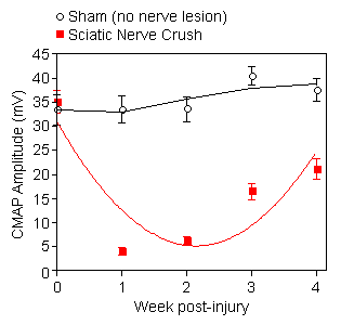
Sciatic nerve crush injury in the mouse is a widely used model for peripheral nerve regeneration. It serves to study underlying cellular and molecular events in nerve injury, and to evaluate therapeutic intervention that might boost axonal regeneration.
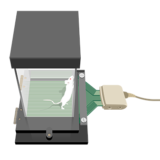
In order to acquire full spectrum of recovery, NEUROFIT monitors both gait and nerve function together with their axon morphology correlates. This model has been tailored for compound testing.
-
Compound testing
Compound testing addresses the effect of chronic treatment of mice (typically starting the same day as the nerve crush injury) on: ☐ recovery of nerve function☐ axonal regeneration
-
Endpoints
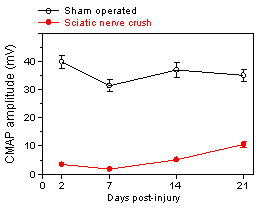
☐ Electrodiagnostic ☐ CMAP ☐ Gait analysis ☐ Nerve morphometry
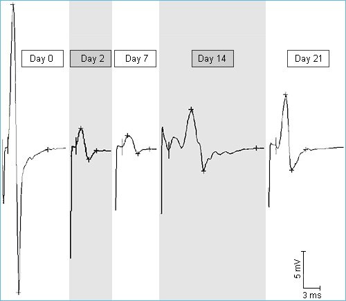
 Electromyography
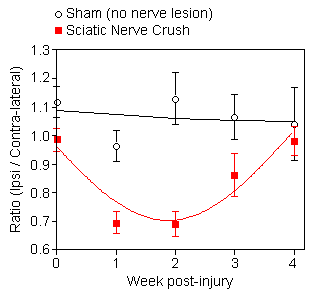
 EMG / BEHAVIOURAL measure
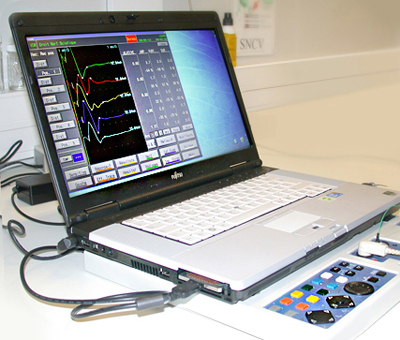
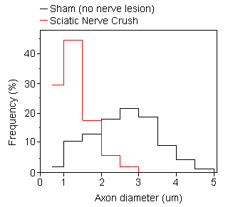
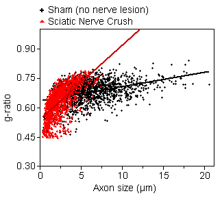
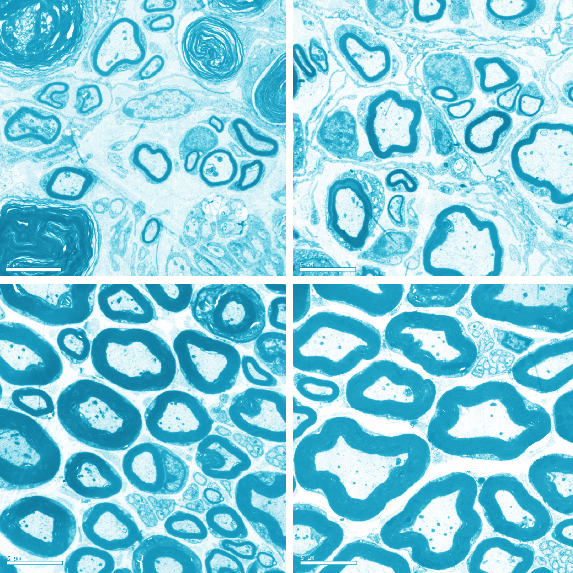
In sham operated specimen, g-ratio (relative thickness of the myelin sheath) is relatively constant for axons of all size. Note the presence of large number of small and hypermyelinated axons following sciatic nerve crush (3 weeks post-injury).

You could also be interested in
-
IENF
The degeneration of intra-epidermal nerve fibers can be monitored and quantified in various animal models of peripheral neuropathy..
Chemotherapy induced neuropathy
Cisplatine, vincristine and taxol are used as antimitotic drugs for treating various cancers. Their employment is often limited by their neurotoxicity.
-
Peripheral neuropathy
Result of damages of the peripheral nervous system. These troubles can be caused either by a trauma or diseases as for example diabetes.
Streptozotocin (STZ)
Diabetic neuropathy can be studied in rat following streptozotocin administration which induces a rapid and persistent hyperglycaemia.